MEIOSIS I
Meiosis is the process by which replicated chromosomes undergo two nuclear divisions to produce four haploid cells, also called meiocytes (sperms and eggs). Diploid (2n) organisms rely on meiosis to produce meiocytes, which have half the ploidy of the parents, for sexual reproduction. Halving the ploidy in meiocytes is essential for restoring the genetic content of the zygote to that of the parents. Meiosis uses similar mechanisms as those employed during mitosis to accomplish the separation and redistribution of chromosomes. However, several features, namely, the pairing and genetic recombination between homologous chromosomes, are unique to meiosis.
The steps leading up to meiosis are similar to those of mitosis – the centrioles and chromosomes are replicated. The amount of DNA in the cell has doubled, and the ploidy of the cell remains the same as before, at 2n. In meiosis I, the phases are analogous to mitosis: prophase I, metaphase I, anaphase I, and telophase I (below figure). Meiosis I proceeds directly to meiosis II without going through interphase.
Meiosis I is unique in that genetic diversity is generated through crossing over and random positioning of homologous chromosomes (bivalent chromosomes). In addition, in meiosis I, the chromosomal number is reduced from diploid (2n) to haploid (n) during this process. (See figure below, where meiosis I begins with a diploid (2n = 4) cell and ends with two haploid (n = 2) cells.) In humans (2n = 46), who have 23 pairs of chromosomes, the number of chromosomes is reduced by half at the end of meiosis I (n = 23).


Prophase I
During prophase I, chromosomal condensation allows chromosomes to be viewed under the microscope. In late prophase I, homologous chromosomes (also called bivalent chromosomes, or bivalents) pair laterally, or side-by-side. At this time they are said to be in synapsis. During synapsis, crossovers – cross-connections that form from breakage and rejoining between sister chromatids – can occur between the paired bivalents, leading to genetic recombination (exchange of genetic material) between the strands involved. The point where a crossover occurs is called a chiasma (plural chiasmata) (see below figure). In figure below, following crossing over, the blue and red chromosomes, which originally carried AA and aa alleles, respectively, now carry Aa alleles in both chromosomes at the end of prophase I. Note that these bivalents have two chromosomes and four chromatids, with one chromosome originating from each parent.

Metaphase I
In metaphase I, each pair of bivalents (two chromosomes, four chromatids total) align on the metaphase plate. This is different from metaphase in mitosis, where all chromosomes align single file on the metaphase plate. The position of each chromosome in the bivalents is random – either parental homolog can appear on each side. This means that there is a 50-50 chance for the daughter cells to get either the mother’s or father’s homolog for each chromosome (see figure below). As shown in the below figure, during metaphase I, bivalents from either parent can align on either side of the cell. In an organism with two sets of chromosomes, there are four ways in which the chromosomes can be arranged, resulting in differences in chromosomal distribution in daughter cells after meiosis I. (A diploid organism with 2n chromosomes will have 2n possible combinations or ways of arranging its chromosomes during metaphase I.)

In a diploid cell with 2 pairs of chromosomes, there are 4 ways to arrange the chromosomes during metaphase I.
Anaphase I
In anaphase I, homologous chromosomes separate. Homologous chromosomes, each containing two chromatids, move to separate poles. Unlike in mitosis, the centromeres do not split and sister chromatids remain paired in anaphase I.

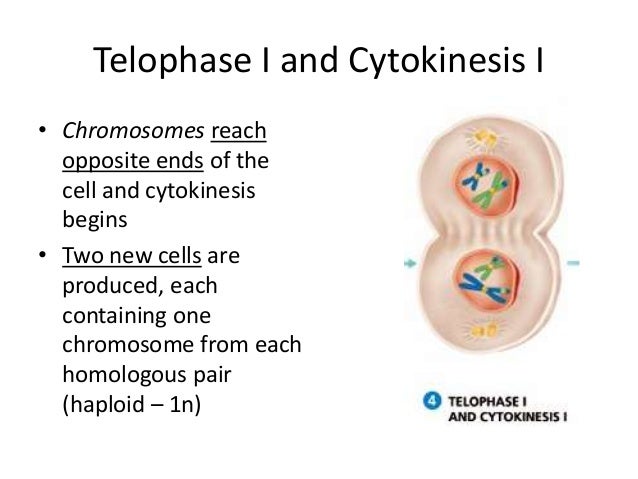
Telophase I and Cytokinesis
In telophase I, the homologs of each bivalent arrive at opposite poles of the cell, and a new nuclear membrane forms around each set of chromosomes. Cytokinesis then divides the cell into two daughter cells. Each of the two daughter cells is now haploid (n), with half the number of chromosomes per nucleus as in meiosis I. In some species, the nuclear membrane briefly forms around the chromosomes, while in others it does not. The cell now proceeds into meiosis II, with the chromosomes remaining condensed.

MEIOSIS II
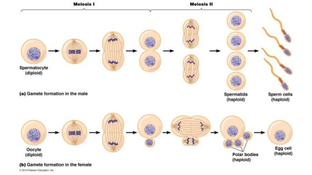
Chromosomal replication does not occur between meiosis I and meiosis II; meiosis I proceeds directly to meiosis II without going through interphase. The second part of the meiosis, meiosis II, resembles mitosis more than meiosis I. Chromosomal numbers, which have already been reduced to haploid (n) by the end of meiosis I, remain unchanged after this division. In meiosis II, the phases are, again, analogous to mitosis: prophase II, metaphase II, anaphase II, and telophase II (see figure below). As shown in the figure below, meiosis II begins with two haploid (n = 2) cells and ends with four haploid (n = 2) cells. Notice that these four meiocytes are genetically different from one another. In humans (2n = 46), who have 23 pairs of chromosomes, the number of chromosomes remains unchanged from the beginning till the end of meiosis II (n = 23).

Prophase II
Spindle fibers reform and attach to centromeres in prophase II.

Metaphase II
The chromosomes align on the metaphase plate during metaphase II in preparation for centromeres to divide in the next phase.

Anaphase II
In anaphase II, chromosomes divide at the centromeres (like in mitosis) and the resulting chromosomes, each with one chromatid, move toward opposite poles of the cell.

Telophase II and Cytokinesis
Four haploid nuclei (containing chromosomes with single chromatids) are formed in telophase II. Division of the cytoplasm during cytokinesis results in four haploid cells. Note that these four cells are not identical, as random arrangements of bivalents and crossing over in meiosis I leads to different genetic composition of these cells.
In humans, meiosis produces genetically different haploid daughter cells, each with 23 chromosomes that consist of one chromatid. These haploid cells become unfertilized eggs in females and sperm in males. The genetic differences ensure siblings of the same parents are never entirely genetically identical.

DNA
Deoxyribonucleic acid or DNA is a molecule that contains the instructions an organism needs to develop, live and reproduce. These instructions are found inside every cell, and are passed down from parents to their children.
DNA structure
DNA is made up of molecules called nucleotides. Each nucleotide contains a phosphate group, a sugar group and a nitrogen base. The four types of nitrogen bases are adenine (A), thymine (T), guanine (G) and cytosine (C). The order of these bases is what determines DNA’s instructions, or genetic code. Human DNA has around 3 billion bases, and more than 99 percent of those bases are the same in all people, according to the U.S. National Library of Medicine (NLM).

Similar to the way the order of letters in the alphabet can be used to form a word, the order of nitrogen bases in a DNA sequence forms genes, which in the language of the cell, tells cells how to make proteins. Another type of nucleic acid, ribonucleic acid, or RNA, translates genetic information from DNA into proteins.
Nucleotides are attached together to form two long strands that spiral to create a structure called a double helix. If you think of the double helix structure as a ladder, the phosphate and sugar molecules would be the sides, while the bases would be the rungs. The bases on one strand pair with the bases on another strand: adenine pairs with thymine, and guanine pairs with cytosine.
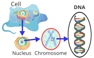
DNA molecules are long — so long, in fact, that they can’t fit into cells without the right packaging. To fit inside cells, DNA is coiled tightly to form structures we call chromosomes. Each chromosome contains a single DNA molecule. Humans have 23 pairs of chromosomes, which are found inside the cell’s nucleus.
DNA discovery
DNA was first observed by a German biochemist named Frederich Miescher in 1869. But for many years, researchers did not realize the importance of this molecule. It was not until 1953 that James Watson, Francis Crick, Maurice Wilkins and Rosalind Franklin figured out the structure of DNA — a double helix — which they realized could carry biological information.
Watson, Crick and Wilkins were awarded the Nobel Prize in Medicine in 1962 “for their discoveries concerning the molecular structure of nucleic acids and its significance for information transfer in living material.” Franklin was not included in the award, although her work was integral to the research. [Related: Unraveling the Human Genome: 6 Molecular Milestones]

DNA sequencing
DNA sequencing is technology that allows researchers to determine the order of bases in a DNA sequence. The technology can be used to determine the order of bases in genes, chromosomes, or an entire genome. In 2000, researchers completed the first full sequence of the human genome, according to a report by the National Human Genome Research Institute.


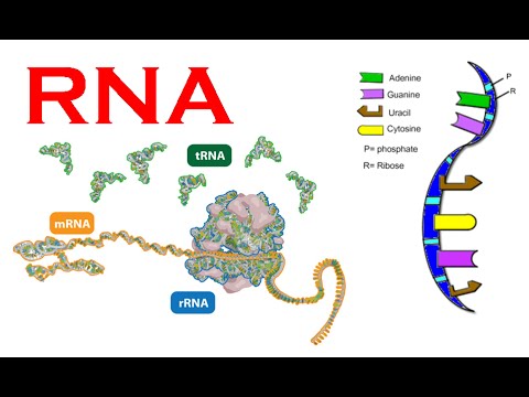
Ribonucleic acid (RNA) is a polymeric molecule essential in various biological roles in coding, decoding, regulation, and expression of genes. RNA and DNA are nucleic acids, and, along with lipids, proteins and carbohydrates, constitute the four major macromolecules essential for all known forms of life. Like DNA, RNA is assembled as a chain of nucleotides, but unlike DNA it is more often found in nature as a single-strand folded onto itself, rather than a paired double-strand. Cellular organisms use messenger RNA (mRNA) to convey genetic information (using the nitrogenous bases guanine, uracil, adenine, and cytosine, denoted by the letters G, U, A, and C) that directs synthesis of specific proteins. Many viruses encode their genetic information using an RNA genome.
There are 4 types of RNA, each encoded by its own type of gene.
The genomic DNA contains all the information for the structure and function of an organism.
In any cell, only some of the genes are expressed, that is, transcribed into RNA.
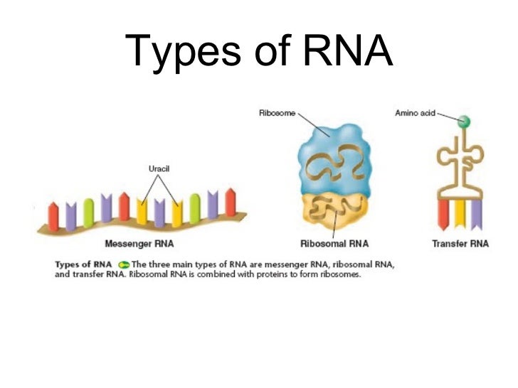
There are 4 types of RNA, each encoded by its own type of gene:
- mRNA – Messenger RNA: Encodes amino acid sequence of a polypeptide.
- tRNA – Transfer RNA: Brings amino acids to ribosomes during translation.
- rRNA – Ribosomal RNA: With ribosomal proteins, makes up the ribosomes, the organelles that translate the mRNA.
- snRNA – Small nuclear RNA: With proteins, forms complexes that are used in RNA processing in eukaryotes. (Not found in prokaryotes.)


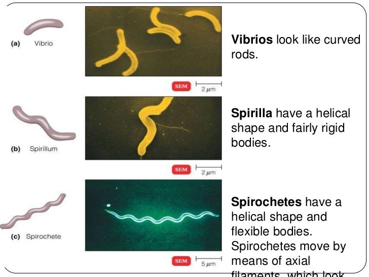 Spirilla (or spirillum for a single cell) are curved bacteria which can range from a gently curved shape to a corkscrew-like spiral. Many spirilla are rigid and capable of movement. A special group of spirilla known as spirochetes are long, slender, and flexible.